Photo: Courtesy: National Human Genome Research Institute (Public domain)
Job description
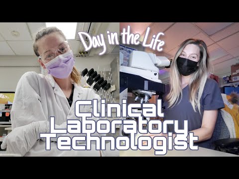
Technologists prepare and view specimens under a microscope, arranging chromosomes into numbered pairs on karyotype charts to spot structural abnormalities that could point to conditions like congenital disorders, fertility problems, or blood disorders. Depending on the case, they choose from techniques like classical cytogenetics, fluorescent in situ hybridization (FISH), or array comparative genome hybridization, running the right instrumentation to produce results a physician can act on. Because those results directly shape a patient's diagnosis or treatment, the work moves fast but leaves little room for error, and it typically requires a Bachelor's degree with specialized lab training.
Pros
- Specialized lab role with a bachelor's degree entry point rather than years of medical school
- Work sits at the center of genetic disease diagnosis, directly informing patient treatment decisions
- Climate-controlled indoor lab setting with a stable, structured daily routine
Cons
- Fast-paced work with constant deadline pressure and quick, high-stakes decisions
- Mistakes in chromosome analysis can have serious consequences for diagnosis and treatment
- Regular exposure to hazardous lab equipment and biological contaminants requires ongoing protective precautions
A typical day
The morning's cases are bone marrow samples from leukemia patients: cells cultured days earlier are harvested, dropped onto slides, and stained so the chromosomes band into their characteristic stripes. At the microscope, the real work begins — counting chromosomes cell by cell, hunting the translocation that confirms a diagnosis, photographing the clearest spreads. Karyotyping follows: arranging chromosome images into their numbered pairs, where a swapped segment between chromosomes 9 and 22 jumps out to a trained eye and changes a patient's treatment plan. Ambiguous findings go to the lab director with notes; clean results get reported to the oncologist the same day. It's meticulous, high-stakes puzzle work — every slide is a person waiting for an answer.
Tasks at the job
- Arrange and attach chromosomes in numbered pairs on karyotype charts, using standard genetics laboratory practices and nomenclature, to identify normal or abnormal chromosomes.
- Count numbers of chromosomes and identify the structural abnormalities by viewing culture slides through microscopes, light microscopes, or photomicroscopes.
- Examine chromosomes found in biological specimens to detect abnormalities.
- Apply prepared specimen and control to appropriate grid, run instrumentation, and produce analyzable results.
- Select appropriate culturing system or procedure based on specimen type and reason for referral.
- Analyze chromosomes found in biological specimens to aid diagnoses and treatments for genetic diseases such as congenital disabilities, fertility problems, and hematological disorders.
Workplace & working conditions
Work happens indoors in a climate-controlled setting, and most of the day is spent sitting at a desk. The role involves regular exposure to hazardous equipment or conditions and contaminants, and standard protective equipment (gloves, safety glasses, and similar) is expected.
The pace is often fast, with real time pressure to meet deadlines, and decisions come up constantly and often need to be made quickly. Mistakes can have serious consequences, so accuracy matters a lot.
Education necessary or common
Bachelor's degree
Common college majors
Licenses & certifications
- ASCP Technologist in Cytogenetics — CG(ASCP)
- State laboratory personnel license where required
Career path
Related fields
Courses to take
- AP Biology — genetics and cell biology are the job's foundation
- Chemistry — staining and lab technique
- Anatomy & Physiology — clinical context for findings
- AP Statistics — quality control and analysis standards
- Health Science/medical terminology electives
Clubs, activities & competitions
- HOSA — medical lab and biotechnology events
- Science Olympiad — designer genes and cell biology events
- Science fair with a genetics project
- Hospital or lab volunteering
First steps you can take now
- Take a free online genetics course and learn what a karyotype shows
- Look at real karyotype images online and practice spotting trisomy 21
- Ask a hospital lab about shadowing a technologist
- Search accredited cytogenetic technologist programs to see the path